Successful Panoramic Radiography at ADK Hospital
N. Vijayan, Radiographer
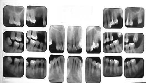
.png) Dental x-rays are pictures of the teeth, bones, and surrounding soft tissues to screen for and help identify problems with the teeth, mouth, and jaw. X-ray pictures can show cavities, hidden dental structures (such as wisdom teeth) and bone loss that cannot be seen during a visual examination. Dental x-rays may also be done as follow up after dental treatments.
Dental x-rays are pictures of the teeth, bones, and surrounding soft tissues to screen for and help identify problems with the teeth, mouth, and jaw. X-ray pictures can show cavities, hidden dental structures (such as wisdom teeth) and bone loss that cannot be seen during a visual examination. Dental x-rays may also be done as follow up after dental treatments.
Panoramic radiography is a specialized extra oral radiographic technique used to examine the upper and the lower jaws in a single film, Panoramic Radiography is also called as Rotational Panoramic Radiography or Pantomography. In this technique, the film and the tube head (X-Ray source) rotate around the patient who remains stationary and produce a series of individual images successively in a single film as these images are combined in a single film overall view of the maxilla, the mandible, and related structures is obtained.
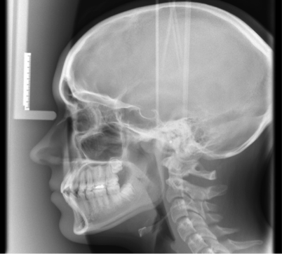
CEPHALOMETRIC RADIOGRAPH
 Radiograph of the head, including the mandible in full lateral view, on tracings of these films, anatomic points, planes, and angles are drawn that assist in the evaluation of the patients' facial growth and development.
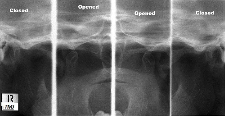
TEMPROMANDIBULAR JOINT
 Tempro mandibular joint (TMJ) syndrome is pain in the jaw joint that can be caused by a variety of medical problems. the TMJ connects the lower jaw (Mandible) to the skull (Temporal bone) in front of the ear ,certain facial muscles control chewing. Problems in this area can cause head and neck pain, facial pain and ear pain.
 The Dental Department at ADK Hospital has been the only facility offering this service in the country. For the past many years, the Hospital has successfully diagnosed problems with the dental and facial bones through this useful technique. Given the success of these imaging investigations, the Hospital is now in the process of introducing the most modern and shophisticated machinery which will immensly benefit the patients. The new installation will give the benefit of digital outputs, the will help improve the quality of the final images and will also allow the radiographers to assit clinicians by image enhancements.

Comments
No comments